Torn Retinaculum Knee : Scarred Ligaments and Retinacula: What does Scar tissue ... / The surgical technique achieved reinforced reattachment of the torn region of the medial retinaculum for improved patellar support and stabilization.
byAdmin•
0
Torn Retinaculum Knee : Scarred Ligaments and Retinacula: What does Scar tissue ... / The surgical technique achieved reinforced reattachment of the torn region of the medial retinaculum for improved patellar support and stabilization.. It thickens as it inserts onto the. It plays important roles in the formation of the fibrous capsule of the knee and in the extension of the knee joint. 1 doctor answer • 1 doctor weighed in. Medial means extending toward the middle. The deep layer is comprised of the lateral patellofemoral ligament, patellotibial band and transverse ligament.
May be the source or a contributing factor of patella femoral pain syndrome (pfps), repetitive or severe injury may lead to stretched/weakened or torn medial retinaculum (dixit). The medial patellar retinaculum is a tendon of the knee that crosses the knee joint on the medial side of the patella. After er doc's saying i must have broken some bone/acl/whatever else, the ortho saying he thought i may have torn my quad tendon, i found out it was none of the above. The superficial layer originates from the lowest fibers of the vastus medialis muscle, sartorius and the medial collateral ligament.the deep layer has contributions from the medial patellofemoral ligament and fascial thickenings. The patella is a sesamoid bone.
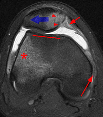
Transient Lateral Patellar Dislocation - Radsource from radsource.us After er doc's saying i must have broken some bone/acl/whatever else, the ortho saying he thought i may have torn my quad tendon, i found out it was none of the above. Lateral trauma to the knee can cause torn medial collateral ligaments, cruciate ligament injury as well as meniscus injury. The medial and lateral patellar retinaculum are on their respective sides of the patella and are continuous with the vastus fascia to the tibia and the patella. My foray into gimp central is due to an apparent knee cap dislocation that has bruised the bone, tore off cartilage from the knee cap, and tore the medial patellar retinaculum. Knee pain is caused by trauma, misalignment, and degeneration as well as by conditions like arthritis. But because of the ligament's location, adequately stretching it can be difficult. The typical injury pattern is a tear of the medial patellofemoral ligament (mpfl) and bone bruises of the patella and the lateral femoral condyle. It plays important roles in the formation of the fibrous capsule of the knee and in the extension of the knee joint.
Lateral trauma to the knee can cause torn medial collateral ligaments, cruciate ligament injury as well as meniscus injury.
The typical injury pattern is a tear of the medial patellofemoral ligament (mpfl) and bone bruises of the patella and the lateral femoral condyle. It thickens as it inserts onto the. The lateral patellar retinaculum is a fibrous expansion comprising of superficial and deep layers. Patellar contusions and avulsion fractures arealsoidentified inupto41% ofpatients i121.thepatellar injuries areadjacent tothe attachment ofthemedial retinaculum and tend toinvolve themedial andinferior aspects ofthebone. The medial patellar retinaculum is a tendon of the knee that crosses the knee joint on the medial side of the patella. Medial means extending toward the middle. The lateral retinaculum is a ligament that helps hold your patella, or kneecap, in place. This retrospective analysis comprised 12 cases of medial retinacular repair in 10 patients. Mri show obliq tear body and posterior horn lateral meniscus, extending infr artic surface and ulceration articular cartilage patella. The medial patellar retinaculum is a fibrous expansion comprising of superficial and deep layers. May be the source or a contributing factor of patella femoral pain syndrome (pfps), repetitive or severe injury may lead to stretched/weakened or torn medial retinaculum (dixit). When the knee moves slightly out of place or becomes tilted in the joint, it can cause tension and pain in the lateral retinaculum. It plays important roles in the formation of the fibrous capsule of the knee and in the extension of the knee joint.
After er doc's saying i must have broken some bone/acl/whatever else, the ortho saying he thought i may have torn my quad tendon, i found out it was none of the above. May be the source or a contributing factor of patella femoral pain syndrome (pfps), repetitive or severe injury may lead to stretched/weakened or torn medial retinaculum (dixit). Stretching this ligament keeps the patella in place and the ligament healthy.stretching a lateral retinaculum of the knee. The medial patellar retinaculum is a tendon of the knee that crosses the knee joint on the medial side of the patella. It thickens as it inserts onto the.
Mpfl ligamentti — the mpfl can tear because of two ... from antwortenkriege.com Mri show obliq tear body and posterior horn lateral meniscus, extending infr artic surface and ulceration articular cartilage patella. Patellar contusions and avulsion fractures arealsoidentified inupto41% ofpatients i121.thepatellar injuries areadjacent tothe attachment ofthemedial retinaculum and tend toinvolve themedial andinferior aspects ofthebone. May be the source or a contributing factor of patella femoral pain syndrome (pfps), repetitive or severe injury may lead to stretched/weakened or torn medial retinaculum (dixit). The first step in treating a torn meniscus is getting the injury examined by a physician who specializes in orthopedics. The typical injury pattern is a tear of the medial patellofemoral ligament (mpfl) and bone bruises of the patella and the lateral femoral condyle. The medial retinaculum plays a minor role — along with the vastus medialis oblique and the medial patellofemoral ligament — in providing medial stability in the knee, according to dr. The patellar tendon often tears at the place where it attaches to the kneecap, and a piece of bone can break off along with the tendon. The lateral retinaculum is a ligament that helps hold your patella, or kneecap, in place.
Knee pain is caused by trauma, misalignment, and degeneration as well as by conditions like arthritis.
The first step in treating a torn meniscus is getting the injury examined by a physician who specializes in orthopedics. 1 doctor answer • 1 doctor weighed in. Mpfl reconstruction is a surgery in which a new medial patellofemoral ligament is created to stabilize the knee and help protect the joint from additional damage. This medical condition requires immediate intervention from an orthopedic surgeon, as sometimes a piece of the patella bone may break off along with the tendon and cannot reattach on its own. The procedure is relatively new. It thickens as it inserts onto the. The medial and lateral patellar retinaculum are on their respective sides of the patella and are continuous with the vastus fascia to the tibia and the patella. The typical injury pattern is a tear of the medial patellofemoral ligament (mpfl) and bone bruises of the patella and the lateral femoral condyle. Additionally, complex injuries to bone, cartilage, and ligaments may occur. They are minor patellar stabilizers and, if intact, can provide knee extension and straight leg raising despite a patellar or quadriceps tendon rupture. When a tear is caused by a medical condition — like tendinitis — the tear usually occurs in the middle of the tendon. Knee pain is caused by trauma, misalignment, and degeneration as well as by conditions like arthritis. When the knee moves slightly out of place or becomes tilted in the joint, it can cause tension and pain in the lateral retinaculum.
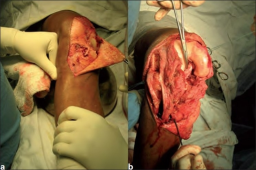
Knee pain is caused by trauma, misalignment, and degeneration as well as by conditions like arthritis. It offers an excellent treatment option for people who have experienced more than one dislocation. The surgical technique achieved reinforced reattachment of the torn region of the medial retinaculum for improved patellar support and stabilization. Medial means extending toward the middle. The medial patellar retinaculum is a tendon of the knee that crosses the knee joint on the medial side of the patella.
Peroperative photograph showing (a) the medial femoral ... from openi.nlm.nih.gov It inserts onto the medial aspect of the patellar. The superficial layer originates from the lowest fibers of the vastus medialis muscle, sartorius and the medial collateral ligament.the deep layer has contributions from the medial patellofemoral ligament and fascial thickenings. It offers an excellent treatment option for people who have experienced more than one dislocation. The patella is a sesamoid bone. Does a high grade partial tear or the medial retinaculum in the knee require surgery or is physical therapy a better option? The injury usually involves an awkward landing from a jumping position where the quadriceps muscle is contracting, but the knee is being forcefully straightened. The medial and lateral patellar retinaculum are on their respective sides of the patella and are continuous with the vastus fascia to the tibia and the patella. It thickens as it inserts onto the.
My foray into gimp central is due to an apparent knee cap dislocation that has bruised the bone, tore off cartilage from the knee cap, and tore the medial patellar retinaculum.
When the knee moves slightly out of place or becomes tilted in the joint, it can cause tension and pain in the lateral retinaculum. They are minor patellar stabilizers and, if intact, can provide knee extension and straight leg raising despite a patellar or quadriceps tendon rupture. The deep layer is comprised of the lateral patellofemoral ligament, patellotibial band and transverse ligament. After er doc's saying i must have broken some bone/acl/whatever else, the ortho saying he thought i may have torn my quad tendon, i found out it was none of the above. A new surgical method is introduced offering a less invasive approach to reattach the medial retinaculum following acute patellar dislocation. Upon flexion of the knee, however, a shortened lateral retinaculum will come under excessive stress as the patella is drawn in the trochlea and the iliotibial band pulls posteriorly on the already shortened lateral retinaculum. This retrospective analysis comprised 12 cases of medial retinacular repair in 10 patients. Stephen pribut's running injuries website. The lateral patellar retinaculum is a fibrous expansion comprising of superficial and deep layers. The superficial layer originates from the lowest fibers of the iliotibial band and from an extension of vastus lateralis fascia. When a tear is caused by a medical condition — like tendinitis — the tear usually occurs in the middle of the tendon. Stretching this ligament keeps the patella in place and the ligament healthy. The patella is a sesamoid bone.
The lateral retinaculum is a ligament that helps hold your patella, or kneecap, in place torn retina. The injury usually involves an awkward landing from a jumping position where the quadriceps muscle is contracting, but the knee is being forcefully straightened.